


Volume 30, No.1 Pages 39 - 42
2. 研究会等報告/WORKSHOP AND COMMITTEE REPORT
The 18th conference of the Asian Crystallographic Association
(公財)高輝度光科学研究センター 研究プロジェクト推進室 生命科学・創薬研究支援基盤グループ Life Science and Drug Discovery Group, Research Project Division, JASRI
1. Introduction
COVID delayed the 2020 conference of the Asian Crystallographic Association (AsCA), which eventually took place at the Kuala Lumpur Convention Centre, Malaysia from 1 to 6 December 2024. The main conference (December 3-6) covered powder X-ray diffraction, single molecule X-ray diffraction and protein crystallography. The theme was "Crystallography today: Beyond the fundamental science". Less fundamental crystallography and more struc-ture analysis, cryo electron microscopy (cryo-EM) and AI. I will mainly discuss the structural biology talks.
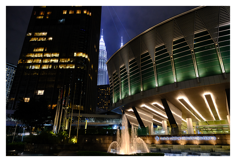
Figure 1 The Kuala Lumpur Convention Centre entrance
Concerning AI, macromolecular databases like https://alphafold.ebi.ac.uk, provide machine-learning Alpha-Fold-generated structures for the complete UniProt database. AI-derived structures can be used to study biological processes, without having to go through the difficult process of solving a structure. Experimental structures are always more reliable than AI-derived models, but generated models can be verified experimentally. This is of interest to laboratories that do not have access to structure-determination facilities, though initiatives like BINDS help remedy that (www.binds.jp). If experimental structural data is available, theoretical models can be fitted into the density, which is helpful if cryo-EM ab initio modelling is difficult in case of ambiguity or low resolution. Furthermore, theoretical models can be used for X-ray molecular replacement.
Single-particle cryo-electron microscopy has been around for a long time, but since the "resolution revolution", this technique has seen great improvements. Proteins are vitrified in water in random orientations. Images are collected of the macromolecules using state-of-the art electron-counting detectors, which record movies that allow correction of beam-induced movement and removal of damaged frames. Individual particles are aligned and averaged in 3D, which results in an electron density-like coulomb potential map in which protein structures can be fitted. Resolutions of macromolecules have been reported up to 1.2 Å, something before never believed possible. Molecular weight is an important factor in the success of 3D single-particle reconstruction, but the limit keeps moving.
2. Lectures Day 1
The opening plenary lecture was by Prof. Megan Maher from The University of Melbourne, Australia. Her group focuses on metal transport and homeostasis in mitochondria.
The keynote speech after the break was by Prof. Chun-Jung Chen at the National Synchrotron Radiation Research Center (NSRRC) in Taiwan. His research group studies viral capsids and assembly. Recent work includes honeybee-infecting Lake Sinai virus, which could be one of the culprits in the Colony Collapse Disorder, which has a big environmental impact. Crystallization of these virus particles was difficult, eventually they used cryo-EM. Interestingly, they found out that the sample consisted of a mixture of different icosahedral particles. This polymorphism could inhibit crystallization, but in cryo-EM they classify into different particles. It is likely that in the absence of genomic material, i.e., heterologous expression of "ghost particles", there is no nucleotide backbone for the capsid proteins to assemble upon, which results in more than one state. Attachment to the correct genome size enforces a larger radius of the particle (49 nm, not 45 nm), capsids contained endogenous DNA.
The group of Prof. Hiroyoshi Matsumura from Ritsumeikan University in Shiga, Japan studies the cell division protein FtsZ, which is a tubulin homolog GTPase. The data resolution was limiting due to its structural heterogeneity. They generated FN3-based monobodies, antibody-like structures. Surface loops are diversified, using DNA techniques, followed by phage-display and selection[1][1] J. Wojcik, O. Hantschel, F. Grebien, I. Kaupe et al.: Nat. Struct. Mol. Biol. 17 (2010) 519.. Monobodies with a specificity towards the protein-protein binding interface on the assembled FtsZ filament were selected, produced, purified and added to the protein sample prior to structure analysis (X-ray/EM). The introduction of these monobodies increased homogeneity and stability, which improved data quality.
I discussed spectroscopic data of frozen crystals, from which diffraction data was collected[2][2] M. Bokhove, T. Kawamura, H. Okumura, S. Goto et al.: J. Biol. Chem. 300 (2024) 107289.. The off-line spec-trometer is near BL26 and on-line at BL41XU, SPring-8.
The afternoon session continued with a keynote lecture by Prof. Eriko Nango from Tohoku University, Miyagi, Japan. She discussed the newly built NanoTerasu synchrotron in Sendai, Japan's first fourth-generation synchrotron radiation facility (nanoterasu.jp). Her talk continued with her group's research on time-resolved protein dynamics using the SACLA X-ray free electron laser (XFEL) facility at SPring-8. She introduced the possibility to study protein dynamics on the nano–to milliseconds timescale. SACLA has a new tape drive system allowing perpendicular X-ray exposure of mixed droplets of microcrystal/reactant for the collection of time resolved data.
Dr. Jaehyun Park introduced the PAL-XFEL facility in Pohang, South Korea, which has a soft and a hard X-ray beam line. In recent years some of the PAL-XFEL microcrystal data was used to design COVID-19 therapeutics. He also presented an apparatus wherein microcrystals are mounted in 2 meters of microtubing in a 2D-array as a static sample device for serial femtosecond X-ray crystallography (SFX) data collection. This mechanism does not allow study of protein dynamics, but it is a low-sample consumption tool to deliver microcrystals to an SFX beam line without using sample streams and keeping crystals in their own mother liquor stable for months, even after X-ray exposure. They now have a more stable microtubing reeling system[3][3] J. Kim, S. Park, Y. Cho, J. Park : Photonics 11 (2024) 95. for SFX.
Prof. Charlie Bond from The University of Western Australia discussed RNA-binding pentatricopeptide repeat (PPR) proteins. An interesting feature of these is that they tend to assemble into infinite helices. However, in the crystals different molecules occupy different asymmetric units (helical disorder). They had to refine the structure as a superposition of different molecules, each with an occupancy of 1/4. Yet, crystals diffracted to 2.0 Å resolution. Upon RNA binding the pitch becomes much shorter, bringing the termini together. They are exploiting this change for the detection of RNA fragments using (single-molecule) Förster resonance energy transfer (FRET).
Eric F. Chen at Thermo Fisher gave a sponsor talk. He discussed their cryo-electron microscopes and application in biology and material science. Recent developments like AI made big leaps, but is still lacking dynamics and ligands. While NMR is a powerful technique, there are some limits due to size and labelling requirements. X-ray crystallography is versatile, but depends on obtaining crystals and unless serial crystallography is used, dynamics are difficult to study. Cryo-EM is becoming more powerful since it can solve structures of different sizes and can be used to study dynamics. Cryo-EM always relied on large molecules; however, recently structures as small as 43 kDa have been solved. ThermoFisher has different cryo-EM platforms and they have aided in the design of new pharmaceutical strategies including drug-antibody adducts, COVID antibodies and tomography. Cryo tomography can be difficult when samples are thick, but recently focused ion beam (FIB) milling has been successful in sample thinning and tomographic data collection. This talk was an ad, but interesting and stimulating nonetheless and may be of interest to people that are less familiar with the currently available EM-applications.
3. Lectures Day 2
An interesting special session talk was from Prof. Horst Puschmann from Durham University, UK. He discussed advancements in the field of small-molecule crystallography, starting from Bragg and Dorothy Hodgkin. The first computers, establishment of the CCDC for small-molecule data by Olga Kennard (ccdc.cam.ac.uk), crystallographic software like shelx, improvements using multimodal refinement, wavefunction based methods and recently PhAI, which uses AI to solve the crystallographic phase problem. Prof. Puschmann develops Olex2 (www.olexsys.org/olex2/), which is a software package for the elucidation and refinement of small molecules. He also presented QCR box, which is a new collaborative project to perform quantum crystallography (qubox.org) and announced the charge density meeting in Durham 2025 (icdm10.netlify.app).
The work of Dr. Terukazu Nogi at Yokohama City University, Japan focuses on crystallography and cryo-EM analysis of in-membrane proteases. These enzymes have an interesting mechanism wherein a transmembrane alpha-helix is cleaved by unwinding into a beta-strand, which forms a beta-sheet with the edge strand of the protease.
Prof. Ning Gao at Peking University, China presented their work on the protein matrix network that shapes red blood cells, a pseudo-hexagonal football-net like, flexible mesh. Yet, the authors were able to box out the protein-complex nodes that connect different strands and perform cryo-EM single-particle averaging to 3.2 Å resolution for this 3.6 MDa complex. Structures docked into the map for model building were experimental or from AlphaFold.
Prof Peter Czabotar at The University of Melbourne introduced their lab's design of specific protein-based inhibitors by AI using Rosetta MotifGraft, resulting in binding constants in the nanomolar range, but specificity was improved by yeast display and next-generation sequencing.
Prof. Hui Ying Yang at Singapore University as part of The Energy and Environmental Sustainability (EES) Laboratory gave a plenary talk. This focused on material science and development of new materials for desalination and the production of drinking water and electricity. The goal is net-zero emission and therefore we need new approaches, such as capacitive deinonization (CDI). She discussed CDI, carbon nanofibers, graphene and carbon nanostructures.
Prof. Nei-Li Chan from the National University Taiwan showed an interesting intermolecular interaction between topoisomerase and a ligand, for the design of new drugs against drug-resistant tuberculosis. The interaction was between a halide (Cl) and an alpha-helical backbone carbonyl, called halogen bonding. One would expect Cl to be electronegative and clash; however, Cl on a benzene ring is δ+, making a strong bond with the carbonyl oxygen. An audience member suggested if the benzene is substituted on the meta-position as well, this interaction is stronger, which could be interesting in the design and modelling of novel ligand-target interactions.
4. Lectures Day 3
Eight young researchers received the Rising Star Award, and gave talks. Hema Kuntrapakam, at the City University New York, USA showed that artificial tripeptides can assemble into complex structures and the order of the amino acids affects their assembly pattern, which I thought interesting.
A sponsored talk of Rigaku by Dr. Christian Göb, Neu-Isenburg, Germany, showed their novel instrument with JEOL, Japan to perform micro-ED. 160º of diffraction data can be collected from (frozen) nm-µm crystals.
Dr. Cong Liu at the Chinese Academy of Sciences, Shanghai gave a keynote talk about structures of amyloid fibres. These protein aggregates in the brain are associated with neurodegenerative diseases such as Huntington's, Alzheimer's and Parkinson's. Different types of assemblies are linked to different pathologies. Alpha-synuclein (Parkinson's) is one of their targets, which they study using cryo-EM helical reconstruction. Amyloids display interesting characteristics such as twisting networks of side chain amide interaction (Asn/Gln) or stacking of aromatic side chains (Tyr/Phe/Trp) across a whole fibre. They design small molecules to break up these interactions, preventing filament growth, which could lead to new drugs.
Day 3 concluded with the conference dinner on the top of the KL tower (282 m). With nice food and cultural dances.
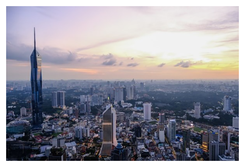
Figure 2 View over Kuala Lumpur city from the KL tower.
5. Lectures Day 4 (last day)
An interesting talk was by the invited speaker Prof. Leonard Chavas at Nagoya University, Japan. His lab has an interesting approach to protein crystallization, in vivo macromolecular crystallography (ivMX). This is challenging; however, one way is to overexpress and target the protein to specific subcellular compartment, e.g., nucleus, cytoplasm, peroxisome, etc. Inside an organelle the sample may be pure and concentrated enough to yield crystals. Crystals are small, but data can be collected using micro- focus serial in vivo crystallography or micro-ED. The ivMX platform is now open to proposals on BINDS[4][4] L. M. G. Chavas, F. Coulibaly, D. Garriga : IUCrJ 11 (2024) 476..
Prof. Fasséli Coulibaly at Monash University, Australia gave a keynote talk entitled "Crystallogenesis in the age of AI". AI can generate any structure; however, certain post-translational modifications cannot be predicted by AI, showing that experimental techniques are important. He also mentioned some methods that can help improve the chance of getting diffraction data such as serial crystallography, microcrystal electron diffraction, in chip diffraction, use of dynamic light scattering crystallisation devices and FIB milling lipid cubic phase-embedded crystals for cryo-EM/ED. Also, in their lab they are doing in vivo crystallography in e.g., silkworm. Methods to overcome the difficulties in detecting microcrystals include non-linear imaging second harmonic microscopy, on-mesh trypan blue-dying and using cell sorters to scan for microcrystals.
The final plenary talk was by Prof. Genji Kurisu at Osaka University, Japan, who is the head the PDBj. The PDB has been shown to be a ground-breaking institution since it was launched in 1971. And no one at the time could have imagined the impact it would have. Deep-learning structure prediction would not have been possible. PDBj, which takes care of all PDB depositions in Asia, is the Japanese arm of the worldwide protein data bank (wwPDB), an offshoot connected to the same infrastructure as the PDB in the USA, the PDBe in Europe and the latest addition the Chinese PDB. There are 228,677 structures, proven invaluable in fundamental biology, biomedicine, bioenergy and more. The PDB contains coordinates and data obtained with NMR, protein crystallography and electron microscopy.The OneDep system reduces biocurator workload and allows a validator to process a 1000 structures per year, resulting in the deposition of fully validated structural data. Many journals require a PDB validation report when submitting a manuscript. New features include updating coordinates while retaining the original ID. Structural data has been proven invaluable during the COVID-19 pandemic. The PDB allowed structures to be deposited while still undergoing validation and optimisation giving researchers a head start to design drugs, antibodies, etc. The wwPDB sees 2 million downloads per day and it is now also on the amazon cloud infrastructure. It is now also possible to deposit raw X-ray data (xrda.pdbj.org) similar to electron microscopy images (ebi.ac.uk/empiar/). The wwPDB celebrated its 53rd birthday in October, this year and Prof. Kurisu's talk showed how valuable the wwPDB is and that it will be around for many years to come.
This concludes AsCA. AsCA2025 will take place in Taiwan with a workshop in Okinawa (AsCA2025.org).
References
[1] J. Wojcik, O. Hantschel, F. Grebien, I. Kaupe et al.: Nat. Struct. Mol. Biol. 17 (2010) 519.
[2] M. Bokhove, T. Kawamura, H. Okumura, S. Goto et al.: J. Biol. Chem. 300 (2024) 107289.
[3] J. Kim, S. Park, Y. Cho, J. Park : Photonics 11 (2024) 95.
[4] L. M. G. Chavas, F. Coulibaly, D. Garriga : IUCrJ 11 (2024) 476.
(公財)高輝度光科学研究センター 研究プロジェクト推進室
生命科学・創薬研究支援基盤グループ
〒679-5198 兵庫県佐用郡佐用町光都1-1-1
e-mail : mbokhove@spring8.or.jp


